Those Crazy Geniuses Who Gave Us MRI: The REAL Story
Table of Contents
How Those Giant Tube Machines Actually Happened
So I was lying in this claustrophobic tube last month getting my knee checked out (stupid soccer injury!) when I started wondering – who the heck came up with this thing(MRI Machine) anyway? Turns out, it’s a pretty wild story about a bunch of science nerds who probably had NO IDEA how many millions of lives they’d end up saving.
Magnetic imaging machines are seriously mind-blowing when you think about it. Doctors can literally see INSIDE your body without cutting you open or blasting you with radiation. Before these machines came along, your doc might’ve had to do exploratory surgery just to figure out what was wrong with you. Yikes!
I’ve done a ton of research on this (cuz I’m kinda weird that way lol) and honestly, the whole story is way more interesting than those boring science textbooks would have you believe.
The Brain Trust Behind the Machine
A bunch of super-smart people contributed to this technology over decades on MRI Machine. It wasn’t just ONE genius in a lab coat shouting “Eureka!” – though that would make a better movie, wouldn’t it?
Isidor Rabi: The Guy Who Started It All (Kinda)
Back in the 1930s (when my grandpa was still a kid!), this physicist named Isidor Rabi noticed something bizarre – atoms would do this weird dance when you put them in a magnetic field and hit them with radio waves.
Rabi wasn’t thinking about medical stuff AT ALL. Dude was just playing around with atoms and magnetic fields because… well, that’s what physicists did back then. They had no Netflix, I guess?
His discovery was super technical (something about “nuclear magnetic resonance”), but basically, he found out that atoms give off detectable signals when you mess with them using magnets. He probably had zero clue this would someday help diagnose my torn meniscus!
Felix Bloch & Edward Purcell: The Dynamic Duo
Fast forward to the 1940s. World War 2 is going on, and two scientists working separately (Bloch at Stanford and Purcell at Harvard) both figured out better ways to detect those magnetic signals Rabi discovered.
These guys refined the whole process and made it WAY more sensitive. They could now see these signals in liquids and solids, not just gas atoms. I’m not gonna pretend I understand all the physics (barely passed that class in high school, tbh), but their work was apparently good enough to win the Nobel Prize in 1952!
The crazy part? They STILL weren’t thinking about medical imaging. They were just physics nerds doing physics stuff. It’s kinda like how people invented the internet to share research papers, and now we use it to watch cat videos and argue with strangers.
Paul Lauterbur: The “Wait, We Could Make PICTURES With This?” Guy
OK so here’s where things get seriously cool. In 1973, this chemist named Paul Lauterbur had what I can only imagine was an actual lightbulb-over-the-head moment. He realized: “Hey, if we carefully control the magnetic field in different areas, we could figure out EXACTLY where the signals are coming from!”
This was the game-changer, folks. Suddenly you could create IMAGES from these signals – not just detect that they existed. Lauterbur basically invented the idea of using magnetic field gradients (fancy term, I know) to create 2D images of stuff.
Legend has it that Lauterbur scribbled his idea on a napkin while eating a hamburger. No clue if that’s actually true, but I choose to believe it because it’s awesome. Some of the best ideas happen over fast food, don’t they?
Peter Mansfield: The “Let’s Make This Actually Useful” Dude
So Lauterbur figured out how to create images, but there was still a HUGE problem – early scans took FOREVER. Like, hours of lying completely still. Not exactly practical when you’ve got fidgety patients and busy hospitals.
Enter Peter Mansfield, a physicist from England. This guy developed crazy-complicated math techniques (my brain hurts just thinking about it) that made the whole process MUCH faster. He also helped create something called “echo-planar imaging” which sounds made-up but apparently was a massive breakthrough.
Thanks to Mansfield, scans that used to take hours could now be done in minutes. As someone who could barely stay still for 30 minutes during my knee scan, I personally thank you, Sir Peter! (Yeah, he got knighted, that’s how big a deal this was).
How This Changed Everything for Docs & Patients
Before these MRI machines, doctors mainly relied on X-rays and CT scans. Those work OK for some things (especially bones), but they use radiation, which isn’t great if you need multiple scans. Plus they suck at showing soft tissues like your brain, muscles, and organs.
My cousin’s a radiologist (the doctors who read these scans), and she’s always going on about how these machines revolutionized medicine. She can see tiny tears in ligaments, early-stage tumors, subtle brain abnormalities – stuff that would’ve been impossible to detect before.
And get this – all without a SINGLE dose of radiation! Just magnets and radio waves! How wild is that?? It’s like something out of Star Trek, except it’s real and happening at your local hospital right now.
The Big Breakthroughs Timeline (Simplified Because I’m Not a Historian)
| When | What Happened | Who to Blame |
|---|---|---|
| 1938 | Discovery that atoms do weird things in magnetic fields | Isidor Rabi |
| 1946 | Better ways to measure those weird magnetic behaviors | Bloch & Purcell |
| 1973 | Figuring out how to make actual IMAGES with it | Paul Lauterbur |
| 1975 | Making the scan process WAY faster | Peter Mansfield |
| 1980s | First machines that actually worked in hospitals | Various companies |
All The Stuff That Went Wrong Along the Way
You think creating a revolutionary medical technology is easy? NOPE! These pioneers faced tons of problems:
Early magnetic imagers were ridiculously slow. The first human body scan reportedly took HOURS, and the poor patient had to lie perfectly still the whole time. Can you imagine? I get antsy after 5 minutes!
The images were super blurry at first. Like, worse-than-my-grandma’s-first-flip-phone-camera blurry.
Nobody believed it would work! Lauterbur’s first paper got rejected by the prestigious journal Nature. Ouch! (They probably felt pretty stupid later.)
Early machines were CRAZY expensive and huge. Only big research hospitals could afford them.
The technology was so complicated that it took a while for regular doctors to understand how to use the images.
Without Lauterbur and Mansfield figuring out their imaging tricks and speedups, this technology might’ve stayed stuck in physics labs forever instead of saving lives in hospitals.
Why You Should Actually Care About This Stuff
Next time you’re getting a scan (and if you’re athletic or over 40, chances are you will eventually!), think about all the brilliant scientists whose work made it possible. It’s pretty mind-blowing that a bunch of physicists messing around with magnets and atoms created something that lets doctors see inside your body without cutting you open.
It’s also a reminder that some of the most important discoveries come from basic science with no immediate practical purpose. These guys weren’t trying to invent medical imaging – they were just curious about how atoms behave in magnetic fields. But their curiosity ended up saving countless lives.
I’m not gonna lie, I still get kinda claustrophobic in those machines, but knowing the amazing story behind them makes me appreciate them a lot more. Also, the headphones they give you nowadays make it way more bearable. Pro tip: ask for classic rock. Way better than the weird ambient stuff they usually offer!
How These Magical Machines Changed Medicine Forever
These magnetic scanning machines completely transformed how doctors figure out what’s wrong with you. Before them, medical folks had to either cut you open to see inside (yikes!), or use X-rays, which are basically just shadow pictures that miss a ton of detail and zap you with radiation. Not ideal!
How It’s Totally Different From Other Ways of Peeking Inside You
So here’s the thing about these scanners that blows my mind – they use absolutely ZERO radiation. None! Instead, they use these super-powerful magnets (seriously, they’re so strong they can yank metal objects across the room – my friend who works in radiology has some wild stories!) and radio waves, kinda like what your car stereo picks up.
The machine basically turns your body’s hydrogen atoms (which are EVERYWHERE in your body, btw) into tiny radio transmitters. How crazy is that?? Your own atoms start broadcasting signals that the machine picks up and converts into images. It’s like your body is temporarily its own radio station! WBDY, broadcasting live from your internal organs!
This means doctors can scan you as often as needed without worrying about radiation exposure. My mom had to get multiple scans for her back problems, and this was a huge relief for her.
The Crazy-Good Detail These Machines Can See
Man, the level of detail in these scans is bonkers. Doctors can spot the tiniest abnormalities that would be completely invisible with other techniques.
I remember my neurologist showing me my brain scan after I got this weird recurring headache. He could point out these super tiny blood vessels and even show me exactly which part of my brain was getting less blood flow. Turned out to be nothing serious (thank god!), but the fact that he could see that level of detail WITHOUT CUTTING MY HEAD OPEN?? Mind-blowing!
Where These Machines Really Shine
Brain stuff: These machines are absolute ROCK STARS at showing brain issues. My neighbor’s kid had seizures, and their scanner showed the exact tiny area causing them. Surgeons knew precisely where to focus, and the kid’s been seizure-free for 3 years now!
Joint problems: As a weekend warrior who’s messed up pretty much every joint at some point (getting old sucks, y’all), I can personally attest that these machines see EVERYTHING in your joints. Every torn ligament, worn cartilage, bone bruise – there’s no hiding from the all-seeing magnetic eye!
Heart issues: They’ve got special scans that can actually show your heart BEATING in real time. My uncle needed one after his heart attack, and the doctors could see exactly which parts of his heart muscle were damaged.
Cancer detection: These machines are absolute game-changers for finding tumors. My aunt’s breast cancer was caught super early because her scanner showed a tiny abnormality that mammograms missed entirely.
Gut problems: Got mysterious abdominal pain? These scanners can check out your liver, kidneys, pancreas, and other internal organs in amazing detail. My coworker avoided unnecessary surgery because his scan showed his pancreas inflammation was actually improving without intervention.
The Scanner Olympics: How Different Imaging Methods Stack Up
| Type | Uses Harmful Rays? | What It’s Good For | Where It Falls Short |
|---|---|---|---|
| X-ray | Yep! | Broken bones, chest problems | Terrible for soft tissue – it’s basically just shadows! |
| CT Scan | Lots of em! | Quick trauma images, detailed bone pics | Radiation exposure, not great for soft tissues |
| Magnetic Scanner | Nope! | Soft tissues, brain, joints, organs | Takes forever, super expensive, loud as heck |
| Ultrasound | Nah | Quick looks at babies, organs, blood flow | Can’t see through bone, limited detail |
Cool New Tricks These Machines Keep Learning
The technology keeps getting better, which is pretty rad. They’ve now got:
Functional scanning that can actually show which parts of your brain are ACTIVE during different tasks. Scientists are using this to map brain function in crazy detail. My psych professor showed us these amazing rainbow-colored brain scans of people doing math problems vs. listening to music. So wild!
Special types that track water molecules moving through your brain tissue to map nerve pathways. Neurosurgeons use these to avoid cutting important brain connections during surgery. Imagine having a roadmap of someone’s brain before operating – absolute sci-fi stuff!
These Machines Are Getting Better All The Time
The scanners keep improving in ways that make a huge difference to patients like me. Newer models are:
WAY less noisy (my first scan in the 90s sounded like being inside a jackhammer, my last one was almost bearable)
Much more spacious (great news for claustrophobic folks)
TONS faster (my knee scan took 20 minutes, which would’ve been an hour+ on older machines)
Super sharp images (the detail is insane compared to older scans)
AI-assisted (computers help analyze the images, catching stuff human eyes might miss)
Making the Whole Experience Suck Less for Patients
If you’ve had one of these scans, you know they’re not exactly spa treatments. You’re stuck in a noisy tube staying perfectly still, which isn’t most people’s idea of fun.
But things have improved SO much! My first scan experience in the 90s was torture – cramped, extremely loud, and took forever. My scan last year? Still not my favorite way to spend an afternoon, but so much better! They gave me headphones with my own Spotify playlist, the machine was wider and less claustrophobic, and it took less than half the time.
Plus, these scans sometimes help you avoid WAY more unpleasant procedures. My buddy was convinced he needed knee surgery until his scan showed it was just inflammation that could be treated with physical therapy. No surgery = big win!
Bottom line: these machines have completely changed how doctors diagnose and treat patients. They show incredible detail without radiation risks, helping spot problems earlier and avoid unnecessary procedures. Every time the technology improves, patients benefit – and as someone who’s been that patient multiple times, I’m super grateful to all those science nerds who made it possible!
Major Milestones in Magnetic Imaging Development
The Early Days: How It All Started
Let me take you back to the mid-1900s when all this crazy-cool tech began. It’s wild to think that the machines that have saved so many lives (including my aunt Carol’s, when they found her brain tumor early enough to treat it) started with some physics geeks just messing around with atoms and magnets.
It all kicked off in 1946 when these two scientists, Felix Bloch and Edward Purcell, independently discovered something called nuclear magnetic resonance (NMR). Basically, they found that atomic nuclei absorb and give off electromagnetic energy when placed in magnetic fields. Super technical stuff, but trust me, it was HUGE.
They had NO IDEA this would someday help diagnose everything from torn ACLs to brain tumors. They were just trying to understand how atoms behave! Their discovery was so important they got the Nobel Prize in Physics in 1952. Wonder if they ever realized what they’d started?
For a long time, this was just a neat lab trick used by chemists to figure out what molecules were made of. Nobody was thinking “medical imaging” yet. It’s kinda like how the guys who invented the first computer never imagined we’d one day use similar technology to scroll through TikTok on the toilet. Innovation is weird like that!
From Chemistry Lab to Hospital: The Big Leap
By the 1970s, some really smart folks started wondering if this atomic behavior could somehow create pictures of the human body. Here’s where the story gets REALLY interesting:
In 1971, this doctor named Raymond Damadian published a paper saying he found differences in NMR signals between cancerous and normal tissues. THIS was the lightbulb moment that got people thinking about medical applications. Damadian wasn’t the most humble guy (he later took out full-page newspaper ads when he didn’t get included in a Nobel Prize), but he definitely saw the potential early on!
Then in 1973, Paul Lauterbur had the brilliant idea of adding magnetic field gradients to create actual 2D images. This was MASSIVE – suddenly we went from just detecting signals to creating actual pictures. His first images were just of test tubes of water, but it proved the concept would work!
Right after that, in 1974, Peter Mansfield developed the math that made image creation way faster. Without his contribution, scans would take hours instead of minutes, which would make the technology pretty useless for actual patients. Can you imagine lying completely still for 3 hours for a scan? No thanks!
These breakthroughs turned a physics curiosity into something that could actually help sick people. It’s amazing how many different scientists had to contribute their pieces to the puzzle!
The Brilliant Minds Who Made It Happen
SO many people contributed to developing these machines. Here are just a few of the standouts:
| Who | What They Did | When |
|---|---|---|
| Felix Bloch & Edward Purcell | Discovered how atoms respond to magnetic fields | 1946 |
| Raymond Damadian | Found that cancerous tissues give different signals | 1971 |
| Paul Lauterbur | Figured out how to create actual images | 1973 |
| Peter Mansfield | Made the whole process way faster | 1974 |
I’m probably missing some important contributors (sorry!), but these were the heavy hitters. What’s cool is how they came from different backgrounds – physics, medicine, chemistry – and each brought something essential to the table.
It’s like when my friends and I make tacos – one makes amazing guacamole, another does the perfect spicy beef, I handle the fresh tortillas… separately we’re okay, but together we create something magical! (OK that’s a stupid comparison but you get what I mean!)
The First Hospital Machines: Clunky but Revolutionary
The 1980s (decade of big hair and bigger magnetic scanners!) saw the first machines installed in hospitals. OMG these early scanners were BEASTS – enormous, super expensive, and not very user-friendly. But even with their limitations, doctors immediately saw their incredible value.
Early developments that got us from lab curiosity to useful hospital equipment included:
Better magnet designs that created stronger, more stable magnetic fields (these things generate fields tens of thousands of times stronger than Earth’s natural magnetic field – WILD!)
Superconducting magnets that could maintain these crazy-strong fields without overheating
Improved coils that helped create clearer images (my radiologist friend says coil design is STILL super important in getting good images)
Computer interfaces that turned the raw data into images doctors could actually understand (early computing was primitive by today’s standards, but it was enough!)
These improvements made the machines reliable enough for regular clinical use, helping diagnose countless patients even in their early forms.
Recent Game-Changers Transforming Today’s Scanners
In just the last 10-15 years, these machines have gotten SO much better! Some of the biggest advances include:
High-field scanners (3 Tesla and up) that produce incredibly detailed images. My neighbor’s a neuroradiologist and says the jump from 1.5T to 3T was like going from standard def to 4K TV – you just see SO much more detail.
Functional imaging that shows brain activity in real-time. This is legitimately sci-fi level stuff – actually SEEING which parts of your brain light up when you perform different tasks!
Open-design scanners that are WAY less claustrophobic. As someone who freaked out during my first scan (embarrassing but true), these wider, more open designs are a godsend for anxious patients.
AI assistance that helps identify abnormalities doctors might miss. The machines are literally getting smarter! My doc showed me how the AI highlighted a tiny abnormality in my shoulder scan that could have been easily overlooked.
These advances have made these machines more useful, more comfortable, and more accessible than ever before.
The Ongoing Story of Innovation
Looking at the whole timeline, it’s pretty mind-blowing how far we’ve come – from basic physics experiments to machines that can show your beating heart in real-time from any angle.
Each milestone built on what came before, with scientists and engineers constantly pushing the boundaries of what’s possible. It reminds me of how each iPhone builds on the previous model – except instead of giving us better selfies, these advances are saving lives!
When I had my shoulder scanned last year after a kayaking mishap (note to self: white water and middle age don’t mix well), I was lying in this state-of-the-art machine thinking about all the incremental improvements that made my diagnosis possible. From Bloch and Purcell’s original discovery to the AI assistance identifying my torn labrum, every step in this journey mattered.
These machines will keep evolving and improving, making diagnoses even more accurate and patient experiences more comfortable. Who knows what capabilities they’ll have in another 10 years? I’m kinda excited to find out (though hopefully NOT because I’ve injured something else by then)!
The Science Behind the Scanner: How the Heck Does This Thing Work?
Magnetic Fields: The Scanner’s Secret Sauce
So here’s the deal with these amazing machines – they rely on INSANELY powerful magnets to get images of your insides. We’re talking magnets so strong they could lift a car! When you slide into that tube, you’re entering a magnetic field that’s like 10,000 times stronger than the Earth’s natural magnetism. Pretty wild, right?
The main job of this super-magnet is to line up all the hydrogen atoms in your body. Why hydrogen? Because it’s EVERYWHERE in your body – in water, fat, proteins, you name it. Normally these hydrogen atoms point in random directions, but when the big magnet turns on, they all line up like soldiers at attention.
I remember asking the tech during my lower back scan (stupid gardening injury!) about how strong the magnet was. She told me they have strict protocols about metal objects because the magnet could literally yank metallic items across the room! That’s why they ask all those questions about metal implants and make you remove jewelry before your scan.
Radio Waves: The Conversation With Your Atoms
Once your hydrogen atoms are all lined up from the magnet, the machine sends radio frequency pulses into your body. Kinda like the radio waves that bring you your favorite songs, but these ones are having a conversation with your atoms instead!
These radio waves temporarily knock your hydrogen atoms out of alignment. And when the radio waves stop, the atoms snap back into position, releasing energy as they do. It’s this released energy that the scanner detects.
The cool thing is, different tissues in your body (muscle, fat, bone, etc.) cause the hydrogen atoms to realign at different speeds. So a brain cell’s hydrogen atoms will “sing” a different “song” than a muscle cell’s atoms. This difference is what creates the contrast in the images.
I’m not gonna pretend I understand all the physics here (I barely passed science in high school lol), but I find it AMAZING that we can use invisible magnetic fields and radio waves to see inside someone’s body. It’s practically magic, except it’s real science!
T1 and T2: The Two Different “Songs” Your Tissues Sing
OK so there are actually two main types of signals the machine measures, called T1 and T2. These are super important because they help doctors tell different tissues apart.
T1 (sometimes called “spin-lattice relaxation”): This measures how quickly hydrogen atoms realign with the magnetic field after being knocked out of position. Some tissues realign quickly, others take longer.
T2 (or “spin-spin relaxation”): This measures how quickly the hydrogen atoms start to fall out of sync with each other after being perfectly aligned by the radio pulse.
My cousin’s a radiologist, and she says choosing between T1 and T2 is like choosing between different Instagram filters – each one shows certain features better than others! T1 is great for showing anatomical structure, while T2 really makes fluid and inflammation pop out.
The first time I saw my knee scan, I was amazed at how clearly you could see the different parts – the cartilage, ligaments, even tiny fluid collections were all visible in different shades of gray. The radiologist switched between T1 and T2 views to show me different aspects of my torn meniscus. Pretty cool (though the diagnosis itself sucked, obviously).
Magnetic Gradients: Creating a 3D Map
Here’s where it gets even cooler – the scanner doesn’t just use one big constant magnetic field. It also has smaller, more precise magnets called “gradient coils” that slightly vary the magnetic field in different directions.
These gradients are what allow the machine to know EXACTLY where in your body each signal is coming from. Without them, you’d just get a jumbled mess of signals with no way to create an actual image.
By rapidly switching these gradients on and off, the scanner creates a 3D map of your body, slice by slice. It’s like cutting through a loaf of bread – each slice shows you a cross-section, and together they create the full picture.
These gradient coils are also what cause all that LOUD KNOCKING NOISE during the scan. They’re literally expanding and contracting as they turn on and off super fast. I always think it sounds like someone’s using a jackhammer right next to my head! Thank goodness for those headphones they give you.
Safety and Comfort: What It’s Actually Like
Unlike X-rays or CT scans, magnetic scanners don’t use radiation, which is a huge advantage. You could get scanned every day for years (though why would you want to??) and not worry about radiation exposure.
BUT – and this is important – these powerful magnets can be dangerous around metal objects. That’s why they ask you a million questions before your scan about implants, pacemakers, previous work with metal (like if you’ve ever been a welder), etc. Most modern implants are MRI-safe, but they still need to know exactly what you have.
As for the experience itself… well, it’s not exactly a spa day. You have to lie completely still in a narrow tube while the machine makes incredibly loud banging and knocking noises around you. I’m not claustrophobic, but even I get a little anxious in there.
My tips: close your eyes BEFORE they slide you in, focus on your breathing, and try to enjoy whatever music they pipe through those headphones. My last scan, I requested 80s rock and got through by mentally focusing on the lyrics to “Don’t Stop Believin'” during the loudest parts!
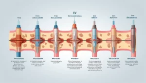
The Step-by-Step Process of Creating an Image
| Stage | What’s Happening | Why It Matters |
|---|---|---|
| Step 1: Alignment | Your hydrogen atoms line up with the big magnet | Creates the baseline state needed for imaging |
| Step 2: Excitation | Radio waves knock atoms out of alignment | Sets up atoms to emit detectable signals |
| Step 3: Relaxation | Atoms release energy as they realign | Different tissues produce different signals |
| Step 4: Detection | Scanner picks up the released energy | Collects the raw data used to build images |
| Step 5: Processing | Computer turns signals into pictures | Creates the images doctors interpret |
Why All This Matters To You
If you’ve ever been nervous about getting scanned (raising my hand here!), understanding how it works might help ease your mind. The whole process is using natural properties of the atoms already in your body – nothing invasive, no radiation, no chemicals being injected (unless you need contrast, which is a whole other thing).
Every time you get a scan, you’re benefiting from decades of scientific breakthroughs that let doctors see inside you without cutting you open. That’s pretty freaking amazing!
And the images these machines produce are INCREDIBLE – detailed enough to show tiny tears in ligaments, early-stage tumors, subtle brain abnormalities, and so much more. My orthopedist was able to see exactly which part of my meniscus was torn and how severe it was, which helped him determine I didn’t need surgery (thank goodness!).
Next time you’re in one of these machines, try to appreciate the mind-boggling science happening all around you – even if it’s hard to feel philosophical when you’re trying not to move for 30 minutes in a noisy tube!
Cutting-Edge Developments Building on the Original Technology
The Never-Ending Evolution
Since they first appeared in hospitals, magnetic scanners have kept improving in ways that make them more useful for patients and doctors. The journey from those early, primitive machines to today’s advanced systems is pretty incredible. The newest innovations are pushing boundaries even further, making scans faster, clearer, and available to more patients than ever before.
My dad had a scan back in the 90s that took almost two hours. Last year, I had a similar scan that took 25 minutes. That’s the kind of practical improvement that makes a HUGE difference in real life!
New Scanning Techniques That Seem Like Science Fiction
The coolest new scanning methods can do things that would’ve seemed impossible even 10 years ago. Functional scanning actually shows brain activity in real-time – doctors can see which parts of your brain “light up” when you perform different tasks. A friend of mine participated in a research study where they could see her brain activity while she solved math problems versus while she listened to music. How crazy is that?!
Another technique called diffusion tensor imaging tracks how water molecules move through brain tissue, creating detailed maps of nerve pathways. This helps neurosurgeons plan operations to avoid cutting critical connections. Before this technology, they were basically going in partially blind and hoping for the best!
Compressed sensing is another game-changer that’s cut scan times dramatically. The machine captures only the essential data needed for diagnosis rather than every possible data point. It’s kind of like when you compress a photo – you lose some information, but not the important stuff. This makes scans WAY more tolerable for fidgety patients (like yours truly – sitting still for an hour? Torture!).
Hardware Upgrades That Make Better Pictures
The machines themselves have gotten seriously upgraded too. Newer scanners use stronger magnets and more sensitive detectors, which means sharper, more detailed images. The jump from 1.5 Tesla to 3 Tesla machines was huge – suddenly doctors could see tiny abnormalities that might have been missed before.
My neighbor’s dad had a small brain aneurysm detected on a 3T scan that had been missed on a previous 1.5T scan. The doctors said the newer machine likely saved his life by catching it before it ruptured. THAT’S the real-world impact of these technical improvements!
There are also portable and lower-field scanning systems being developed to bring this technology to places that can’t afford or accommodate traditional machines. I read about a portable scanner being used after earthquakes to check injured people right at disaster sites. How amazing is that?
How AI is Making These Machines Even Smarter
Artificial intelligence is revolutionizing how these images are analyzed. AI algorithms can detect subtle abnormalities that human eyes might miss, helping with early diagnosis of diseases like cancer or multiple sclerosis.
My radiologist told me they’re now using AI to help analyze spine images, and it’s catching disc herniations and nerve compression that sometimes get overlooked in routine readings. The AI doesn’t replace the doctor – it’s more like having a super-attentive assistant who never gets tired or distracted.
AI can also customize scan settings in real-time based on each patient’s unique anatomy and medical condition, improving both accuracy and speed. The machine actually adapts to YOUR body during the scan!
Making the Experience Better for Patients
Anyone who’s had a scan knows it’s not exactly a fun time. You’re stuck in a tube, trying not to move, while the machine makes incredibly loud noises around you. Thankfully, newer systems focus on improving this experience.
Modern machines are significantly quieter (though still not what I’d call “quiet” lol), and many have wider openings to reduce that claustrophobic feeling. My first scan in the 90s was a nightmare – I almost had a panic attack. My most recent one was almost… bearable? Progress!
Safety measures have also improved dramatically. Better shielding and monitoring reduce risks from the magnetic fields and radio frequency energy. This makes scanning safer for more patients, including children and people with certain types of implants who previously couldn’t be scanned.
The Next Big Things on the Horizon
| The New Tech | What It Could Do | Why You Should Care |
|---|---|---|
| 7 Tesla Scanners | Show brain and joint details we’ve never seen before | Could diagnose conditions earlier when they’re more treatable |
| AI Diagnostic Systems | Automatically flag concerning findings | Faster results and fewer missed diagnoses |
| Portable Systems | Bring scanning to remote locations | Emergency diagnosis when you can’t get to a hospital |
| Ultra-Fast Protocols | Complete comprehensive scans in minutes | Less time trapped in the tube = happier patients! |
Teams Making Tomorrow’s Breakthroughs
The future of scanning technology depends on collaboration between all kinds of experts – physicists figuring out the basic science, engineers building better hardware, computer scientists creating smarter software, and doctors identifying clinical needs.
I had dinner with a friend who works at a research hospital recently, and she told me their scanning research team includes people from SEVEN different scientific fields all working together. That kind of teamwork is what pushes medical technology forward!
What We Can Look Forward To
Despite competition from newer technologies, magnetic scanning remains one of medicine’s most powerful diagnostic tools because it’s non-invasive and gives incredible detail of soft tissues. The innovations building on those original discoveries promise to make scanning even more precise, user-friendly, and accessible.
As these technologies continue to advance, we can expect faster diagnoses, more effective treatment planning, and scanning becoming available to patients worldwide who currently don’t have access. My cousin lives in a rural area and currently has to drive 2+ hours to reach the nearest scanner – but portable or lower-cost systems could eventually bring this technology much closer to home.
While the science can seem complicated (and it is!), the goal is beautifully simple: helping doctors see what’s wrong so they can make us better. And THAT’S something worth investing in!
Last Thoughts
The story of magnetic scanning is honestly one of the coolest examples of human ingenuity I’ve ever come across. What started with some curious physicists playing around with atoms and magnets eventually transformed into technology that saves lives every single day. The brilliant scientists who built this technology piece by piece probably never imagined how many millions of patients would benefit from their work.
The science that makes these machines function – those powerful magnetic fields and precisely tuned radio waves – creates detailed images of our insides that would have seemed like pure science fiction just a few generations ago. Every time I see one of these machines, I’m amazed that we can non-invasively peek inside the human body with such incredible detail. It lets doctors diagnose diseases earlier, plan treatments more precisely, and monitor patients’ progress without additional risk.
Looking ahead, the innovations building on these foundational discoveries promise even more incredible capabilities – faster scans, more detailed images, and accessibility for patients worldwide. From 7-Tesla super-scanners that show microscopic details to portable units that can bring this technology to remote locations, the future of magnetic imaging looks brighter than ever.
For anyone who’s curious about how medical technology evolves, the magnetic scanner’s journey offers powerful lessons about scientific curiosity, perseverance, and collaboration across different fields. By appreciating the brilliant minds behind this technology, we gain perspective on how seemingly abstract scientific discoveries can transform into tools that touch millions of lives.
Next time you or someone you care about needs one of these scans, remember you’re benefiting from one of medicine’s greatest success stories – a perfect example of how human curiosity